6914 results
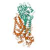
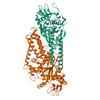
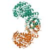
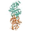
X-ray diffraction data for the Crystal structure of Cysteinyl-tRNA synthetase (CysRS) from Plasmodium falciparum in complex with O5'-(L-GLUTAMYL-SULFAMOYL)-ADENOSINE
First author:
Seattle Structural Genomics Center for Infectious Disease (SSGCID)
Uniprot: Q8IJP3
Resolution: 2.03 Å
R/Rfree: 0.18/0.20
Uniprot: Q8IJP3
Resolution: 2.03 Å
R/Rfree: 0.18/0.20
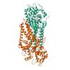
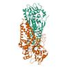
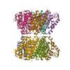
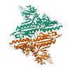
X-ray diffraction data for the Crystal structure of Cysteinyl-tRNA synthetase (CysRS) from Plasmodium falciparum in complex with Cysteine
First author:
Seattle Structural Genomics Center for Infectious Disease (SSGCID)
Uniprot: Q8IJP3
Resolution: 2.16 Å
R/Rfree: 0.19/0.22
Uniprot: Q8IJP3
Resolution: 2.16 Å
R/Rfree: 0.19/0.22
X-ray diffraction data for the Crystal structure of Cysteinyl-tRNA synthetase (CysRS) from Plasmodium falciparum in complex with ADP
First author:
Seattle Structural Genomics Center for Infectious Disease (SSGCID)
Uniprot: Q8IJP3
Resolution: 2.14 Å
R/Rfree: 0.18/0.20
Uniprot: Q8IJP3
Resolution: 2.14 Å
R/Rfree: 0.18/0.20
X-ray diffraction data for the Crystal structure of Cysteinyl-tRNA synthetase (CysRS) from Plasmodium falciparum in complex with AMP and Cysteine
First author:
Seattle Structural Genomics Center for Infectious Disease (SSGCID)
Uniprot: Q8IJP3
Resolution: 2.34 Å
R/Rfree: 0.19/0.22
Uniprot: Q8IJP3
Resolution: 2.34 Å
R/Rfree: 0.19/0.22
X-ray diffraction data for the Crystal structure of a Glyceraldehyde-3-phosphate dehydrogenase from Bordetella pertussis (monoclinic P form)
First author:
Seattle Structural Genomics Center for Infectious Disease (SSGCID) Seattle Structural Genomics Center for Infectious Disease
Uniprot: Q7VZB9
Resolution: 2.00 Å
R/Rfree: 0.17/0.20
Uniprot: Q7VZB9
Resolution: 2.00 Å
R/Rfree: 0.17/0.20
X-ray diffraction data for the Crystal Structure of Acetyl-CoA synthetase from Cryptococcus neoformans H99 in complex with inhibitor HGN-1310 (dd3-027)
First author:
Seattle Structural Genomics Center for Infectious Disease (SSGCID) Seattle Structural Genomics Center for Infectious Disease
Uniprot: J9VFT1
Resolution: 2.40 Å
R/Rfree: 0.18/0.22
Uniprot: J9VFT1
Resolution: 2.40 Å
R/Rfree: 0.18/0.22
X-ray diffraction data for the Rhombohedral crystalline form of human insulin complexed with m-cresol
X-ray diffraction data for the Crystal structure of Apo Cysteinyl-tRNA synthetase (CysRS) from Plasmodium falciparum (Orthrhombic P form)
First author:
Seattle Structural Genomics Center for Infectious Disease (SSGCID)
Uniprot: Q8IJP3
Resolution: 3.06 Å
R/Rfree: 0.23/0.29
Uniprot: Q8IJP3
Resolution: 3.06 Å
R/Rfree: 0.23/0.29
X-ray diffraction data for the Crystal structure of Cysteinyl-tRNA synthetase (CysRS) from Plasmodium falciparum in complex with 5'-Sulfamoyladenosine

First author:
Seattle Structural Genomics Center for Infectious Disease (SSGCID)
Uniprot: Q8IJP3
Resolution: 2.77 Å
R/Rfree: 0.19/0.23
Uniprot: Q8IJP3
Resolution: 2.77 Å
R/Rfree: 0.19/0.23
X-ray diffraction data for the Crystal structure of Cysteinyl-tRNA synthetase (CysRS) from Plasmodium falciparum in complex with cysteinyl-AMP
First author:
Seattle Structural Genomics Center for Infectious Disease (SSGCID)
Uniprot: Q8IJP3
Resolution: 2.22 Å
R/Rfree: 0.18/0.20
Uniprot: Q8IJP3
Resolution: 2.22 Å
R/Rfree: 0.18/0.20
X-ray diffraction data for the Structure of Saro_1862, a UPF0261 domain protein from Novosphingobium aromaticivorans with bound acetovanillone
X-ray diffraction data for the Crystal Structure of 6,7-dimethyl-8-ribityllumazine synthase from Bordetella pertussis in complex with 5-amino-6-(D-ribitylamino)uracil
First author:
Seattle Structural Genomics Center for Infectious Disease (SSGCID)
Uniprot: Q7VTN4
Resolution: 2.56 Å
R/Rfree: 0.20/0.25
Uniprot: Q7VTN4
Resolution: 2.56 Å
R/Rfree: 0.20/0.25
X-ray diffraction data for the Crystal structure of Glutamate--tRNA ligase (GltX) from Moraxella catarrhalis in complex with 5'-O-(N-Glutamyl)sulfamoyladeonosine
First author:
Seattle Structural Genomics Center for Infectious Disease (SSGCID) Seattle Structural Genomics Center for Infectious Disease
Uniprot: A0AB36DQE3
Resolution: 2.92 Å
R/Rfree: 0.21/0.25
Uniprot: A0AB36DQE3
Resolution: 2.92 Å
R/Rfree: 0.21/0.25
X-ray diffraction data for the Crystal structure of Prolyl-tRNA synthetase (ProRS, Proline--tRNA ligase) from Plasmodium falciparum in complex with inhibitor YNW69
First author:
Seattle Structural Genomics Center for Infectious Disease (SSGCID)
Uniprot: Q8I5R7
Resolution: 2.43 Å
R/Rfree: 0.20/0.23
Uniprot: Q8I5R7
Resolution: 2.43 Å
R/Rfree: 0.20/0.23
X-ray diffraction data for the Crystal structure of nucleoside-diphosphate kinase Cryptosporidium parvum (GMP complex)
First author:
Seattle Structural Genomics Center for Infectious Disease (SSGCID) Seattle Structural Genomics Center for Infectious Disease
Uniprot: Q5CR64
Resolution: 1.64 Å
R/Rfree: 0.15/0.18
Uniprot: Q5CR64
Resolution: 1.64 Å
R/Rfree: 0.15/0.18