6890 results
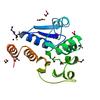
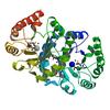
X-ray diffraction data for the Crystal structure of Nitroreductase with Bound FMN (YP_211706.1) from Bacteroides fragilis NCTC 9343 at 1.70 A resolution
First author:
Joint Center for Structural Genomics (JCSG)
Resolution: 1.70 Å
R/Rfree: 0.15/0.18
Resolution: 1.70 Å
R/Rfree: 0.15/0.18
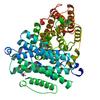
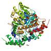
X-ray diffraction data for the Crystal structure of a Nitroreductase with bound FMN (Dhaf_2018) from Desulfitobacterium hafniense DCB-2 at 2.30 A resolution
First author:
Joint Center for Structural Genomics (JCSG)
Resolution: 2.30 Å
R/Rfree: 0.17/0.24
Resolution: 2.30 Å
R/Rfree: 0.17/0.24
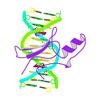
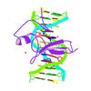
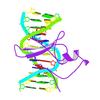
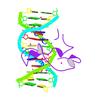
X-ray diffraction data for the Human METTL3-METTL14 complex
First author:
H. ZENG
Resolution: 1.80 Å
R/Rfree: 0.20/0.22
Resolution: 1.80 Å
R/Rfree: 0.20/0.22
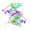
X-ray diffraction data for the Fragment of 7SK snRNA methylphosphate capping enzyme
First author:
H. Wu
Resolution: 2.55 Å
R/Rfree: 0.20/0.22
Resolution: 2.55 Å
R/Rfree: 0.20/0.22
First author:
K. Liu
Resolution: 2.30 Å
R/Rfree: 0.23/0.29
Resolution: 2.30 Å
R/Rfree: 0.23/0.29
X-ray diffraction data for the Crystal structure of predicted HD superfamily hydrolase (104161995) from uncultured Thermotogales bacterium at 1.45 A resolution
First author:
Joint Center for Structural Genomics (JCSG)
Resolution: 1.45 Å
R/Rfree: 0.18/0.19
Resolution: 1.45 Å
R/Rfree: 0.18/0.19
First author:
K. Liu
Resolution: 2.01 Å
R/Rfree: 0.21/0.25
Resolution: 2.01 Å
R/Rfree: 0.21/0.25
First author:
K. Liu
Resolution: 1.90 Å
R/Rfree: 0.22/0.25
Resolution: 1.90 Å
R/Rfree: 0.22/0.25
First author:
K. Liu
Resolution: 1.95 Å
R/Rfree: 0.22/0.26
Resolution: 1.95 Å
R/Rfree: 0.22/0.26
First author:
M. Lei
Resolution: 2.30 Å
R/Rfree: 0.23/0.27
Resolution: 2.30 Å
R/Rfree: 0.23/0.27
First author:
M. Lei
Resolution: 2.30 Å
R/Rfree: 0.23/0.27
Resolution: 2.30 Å
R/Rfree: 0.23/0.27
First author:
M. Lei
Resolution: 1.84 Å
R/Rfree: 0.21/0.25
Resolution: 1.84 Å
R/Rfree: 0.21/0.25
First author:
K. Liu
Resolution: 2.05 Å
R/Rfree: 0.23/0.27
Resolution: 2.05 Å
R/Rfree: 0.23/0.27
First author:
K. Liu
Resolution: 2.65 Å
R/Rfree: 0.21/0.24
Resolution: 2.65 Å
R/Rfree: 0.21/0.24
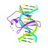
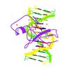
X-ray diffraction data for the MBD2 in complex with methylated DNA
First author:
K. Liu
Resolution: 2.15 Å
R/Rfree: 0.21/0.23
Resolution: 2.15 Å
R/Rfree: 0.21/0.23
X-ray diffraction data for the Crystal structure of MBD2 complex with methylated CpG island
First author:
C. Bian
Resolution: 2.10 Å
R/Rfree: 0.20/0.22
Resolution: 2.10 Å
R/Rfree: 0.20/0.22
First author:
C. Xu
Resolution: 1.80 Å
R/Rfree: 0.23/0.25
Resolution: 1.80 Å
R/Rfree: 0.23/0.25
X-ray diffraction data for the Complex of MBD1-MBD and methylated DNA
First author:
K. Liu
Resolution: 2.25 Å
R/Rfree: 0.24/0.27
Resolution: 2.25 Å
R/Rfree: 0.24/0.27
X-ray diffraction data for the Crystal structure of human monoamine oxidase B (MAO B) in complex with fluorophenyl-chromone-carboxamide
First author:
J. Reis
Resolution: 1.70 Å
R/Rfree: 0.16/0.19
Resolution: 1.70 Å
R/Rfree: 0.16/0.19
X-ray diffraction data for the Crystal structure of human monoamine oxidase B (MAO B) in complex with dimethylphenyl-chromone-carboxamide

First author:
J. Reis
Resolution: 1.80 Å
R/Rfree: 0.17/0.20
Resolution: 1.80 Å
R/Rfree: 0.17/0.20
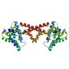
X-ray diffraction data for the Crystal structure of a DJ-1 (PARK7) from Homo sapiens at 1.23 A resolution
First author:
Partnership for Nuclear Receptor Signaling Code Biology (NHRs) Joint Center for Structural Genomics (JCSG)
Resolution: 1.23 Å
R/Rfree: 0.12/0.13
Resolution: 1.23 Å
R/Rfree: 0.12/0.13
X-ray diffraction data for the Crystal structure of a ribonucleotide reductase M2 B (RNRR2) from Homo sapiens at 2.20 A resolution
First author:
Partnership for T-Cell Biology (TCELL) Joint Center for Structural Genomics (JCSG)
Resolution: 2.20 Å
R/Rfree: 0.19/0.22
Resolution: 2.20 Å
R/Rfree: 0.19/0.22
X-ray diffraction data for the Crystal structure of a D-ribulose-5-phosphate-3-epimerase (NP_954699) from HOMO SAPIENS at 2.20 A resolution
First author:
Joint Center for Structural Genomics (JCSG)
Resolution: 2.20 Å
R/Rfree: 0.16/0.21
Resolution: 2.20 Å
R/Rfree: 0.16/0.21
X-ray diffraction data for the Crystal structure of Putative aminotransferase (AAH25799.1) from MUS MUSCULUS at 1.80 A resolution
First author:
Joint Center for Structural Genomics (JCSG)
Resolution: 1.80 Å
R/Rfree: 0.17/0.21
Resolution: 1.80 Å
R/Rfree: 0.17/0.21
X-ray diffraction data for the Crystal structure of Putative aminotransferase (AAH25799.1) from MUS MUSCULUS at 1.65 A resolution
First author:
Joint Center for Structural Genomics (JCSG)
Resolution: 1.65 Å
R/Rfree: 0.17/0.20
Resolution: 1.65 Å
R/Rfree: 0.17/0.20