6907 results
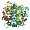
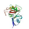
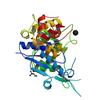
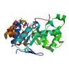
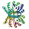
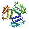
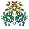
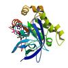
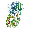
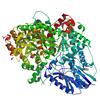
X-ray diffraction data for the Crystal structure of beta-hexosaminidase 1 from Burkholderia cenocepacia J2315 with bound N-Acetyl-D-Glucosamine
First author:
D.R. Davies
Resolution: 1.50 Å
R/Rfree: 0.15/0.18
Resolution: 1.50 Å
R/Rfree: 0.15/0.18
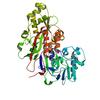
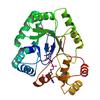
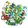
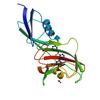
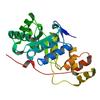
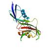
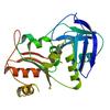
X-ray diffraction data for the X-ray crystal structure of a periplasmic oligopeptide-binding protein/Oligopeptide ABC transporter(OppAIV) from Borrelia burgdorferi
First author:
J.W. Fairman
Resolution: 2.20 Å
R/Rfree: 0.17/0.22
Resolution: 2.20 Å
R/Rfree: 0.17/0.22
X-ray diffraction data for the X-ray crystal structure of a hypothetical deoxyuridine 5-triphosphate nucleotidohydrolase from Mycobacterium abscessus


First author:
L. Baugh
Resolution: 1.65 Å
R/Rfree: 0.16/0.18
Resolution: 1.65 Å
R/Rfree: 0.16/0.18
X-ray diffraction data for the Crystal structure of a SMT fusion Peptidyl-prolyl cis-trans isomerase with surface mutation D44G from Burkholderia pseudomallei complexed with CJ183
First author:
D.W. Begley
Resolution: 2.45 Å
R/Rfree: 0.22/0.26
Resolution: 2.45 Å
R/Rfree: 0.22/0.26
X-ray diffraction data for the Crystal structure of prostaglandin F synthase from Trypanosoma cruzi bound to NADP
First author:
S.O. Moen
Resolution: 1.25 Å
R/Rfree: 0.12/0.14
Resolution: 1.25 Å
R/Rfree: 0.12/0.14
X-ray diffraction data for the X-ray Crystal Structure of Gamma-glutamyl phosphate reductase from Burkholderia thailandensis

First author:
L. Baugh
Resolution: 2.25 Å
R/Rfree: 0.18/0.23
Resolution: 2.25 Å
R/Rfree: 0.18/0.23
X-ray diffraction data for the Crystal structure of a SMT fusion Peptidyl-prolyl cis-trans isomerase from Burkholderia pseudomallei complexed with CJ40
First author:
D.W. Begley
Resolution: 1.95 Å
R/Rfree: 0.19/0.22
Resolution: 1.95 Å
R/Rfree: 0.19/0.22
X-ray diffraction data for the Crystal structure of a glutamyl-tRNA synthetase GluRS from Burkholderia thailandensis bound to L-glutamate
First author:
S.O. Moen
Resolution: 2.05 Å
R/Rfree: 0.20/0.24
Resolution: 2.05 Å
R/Rfree: 0.20/0.24
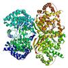
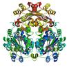
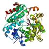
X-ray diffraction data for the Crystal structure of beta-hexosaminidase 1 from Burkholderia cenocepacia J2315
First author:
Davies Seattle Structural Genomics Center for Infectious Disease (SSGCID)
Resolution: 1.38 Å
R/Rfree: 0.16/0.19
Resolution: 1.38 Å
R/Rfree: 0.16/0.19
X-ray diffraction data for the Crystal structure of a COG1565 superfamily member and likely methyl transferase from Burkholderia thailandensis bound to S-adenosyl-homocysteine


First author:
L. Baugh
Resolution: 1.80 Å
R/Rfree: 0.16/0.20
Resolution: 1.80 Å
R/Rfree: 0.16/0.20
X-ray diffraction data for the X-ray crystal structure of Prostaglandin f synthase from Leishmania major Friedlin bound to NADPH
First author:
S.O. Moen
Resolution: 1.80 Å
R/Rfree: 0.17/0.22
Resolution: 1.80 Å
R/Rfree: 0.17/0.22
X-ray diffraction data for the Crystal structure of a SMT fusion Peptidyl-prolyl cis-trans isomerase with surface mutation D44G from Burkholderia pseudomallei complexed with CJ168
First author:
D.W. Begley
Resolution: 1.75 Å
R/Rfree: 0.17/0.22
Resolution: 1.75 Å
R/Rfree: 0.17/0.22
X-ray diffraction data for the Crystal structure of triosephosphate isomerase from Burkholderia thailandensis
First author:
L. Baugh
Resolution: 2.35 Å
R/Rfree: 0.17/0.20
Resolution: 2.35 Å
R/Rfree: 0.17/0.20
X-ray diffraction data for the Crystal structure of prostaglandin F synthase from Trypanosoma cruzi
First author:
S.O. Moen
Resolution: 2.60 Å
R/Rfree: 0.19/0.23
Resolution: 2.60 Å
R/Rfree: 0.19/0.23
X-ray diffraction data for the Crystal Structure of Urease subunit gamma 2 from Brucella melitensis biovar Abortus 2308
First author:
J.W. Fairman
Resolution: 2.10 Å
R/Rfree: 0.15/0.19
Resolution: 2.10 Å
R/Rfree: 0.15/0.19
X-ray diffraction data for the Crystal structure of a SMT fusion Peptidyl-prolyl cis-trans isomerase with surface mutation D44G from Burkholderia pseudomallei complexed with CJ37
First author:
D.W. Begley
Resolution: 1.95 Å
R/Rfree: 0.17/0.22
Resolution: 1.95 Å
R/Rfree: 0.17/0.22
X-ray diffraction data for the Crystal structure of nucleoside diphosphate kinase B from Trypanosoma brucei bound to GTP
First author:
A.S. Gardberg
Resolution: 1.95 Å
R/Rfree: 0.15/0.19
Resolution: 1.95 Å
R/Rfree: 0.15/0.19
X-ray diffraction data for the Crystal structure of nucleoside diphosphate kinase B from Trypanosoma brucei bound to CDP
First author:
A.S. Gardberg
Resolution: 1.70 Å
R/Rfree: 0.15/0.18
Resolution: 1.70 Å
R/Rfree: 0.15/0.18
X-ray diffraction data for the Crystal structure of FERREDOXIN-NADP REDUCTASE from burkholderia thailandensis E264 with bound FAD
First author:
L. Baugh
Resolution: 2.10 Å
R/Rfree: 0.20/0.23
Resolution: 2.10 Å
R/Rfree: 0.20/0.23
X-ray diffraction data for the Crystal structure of the N-terminal domain of Hantaan virus strain 76-118 nucleoprotein
First author:
T.E. Edwards
Resolution: 2.20 Å
R/Rfree: 0.19/0.24
Resolution: 2.20 Å
R/Rfree: 0.19/0.24
X-ray diffraction data for the Crystal structure of a 4-aminobutyrate aminotransferase (GabT) from Mycobacterium abscessus


First author:
L. Baugh
Resolution: 1.80 Å
R/Rfree: 0.14/0.17
Resolution: 1.80 Å
R/Rfree: 0.14/0.17
X-ray diffraction data for the X-ray crystal structure of a putative thioredoxin reductase from Burkholderia cenocepacia
First author:
J.W. Fairman
Resolution: 1.85 Å
R/Rfree: 0.18/0.22
Resolution: 1.85 Å
R/Rfree: 0.18/0.22
X-ray diffraction data for the Crystal structure of ferredoxin-NADP reductase from burkholderia thailandensis E264
First author:
L. Baugh
Resolution: 2.35 Å
R/Rfree: 0.20/0.25
Resolution: 2.35 Å
R/Rfree: 0.20/0.25
X-ray diffraction data for the Crystal structure of a Glutamine dependent NAD+ synthetase from Burkholderia thailandensis
First author:
L. Baugh
Resolution: 1.75 Å
R/Rfree: 0.15/0.18
Resolution: 1.75 Å
R/Rfree: 0.15/0.18
X-ray diffraction data for the X-Ray crystal structure of PLP bound Threonine synthase from Brucella melitensis
First author:
J.S. Christensen
Resolution: 1.90 Å
R/Rfree: 0.18/0.21
Resolution: 1.90 Å
R/Rfree: 0.18/0.21