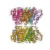
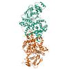
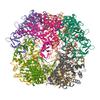
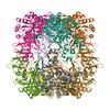
6914 results
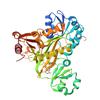
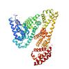
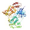
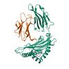
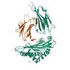
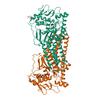
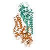
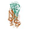
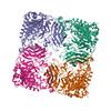
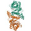
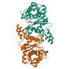
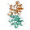
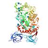
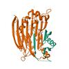
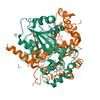
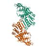
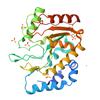
X-ray diffraction data for the Crystal structure of an Iole protein from Brucella melitensis (cobalt complex)
First author:
Seattle Structural Genomics Center for Infectious Disease (SSGCID)
Uniprot: Q8YCG0
Resolution: 2.30 Å
R/Rfree: 0.19/0.24
Uniprot: Q8YCG0
Resolution: 2.30 Å
R/Rfree: 0.19/0.24
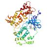
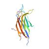
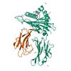
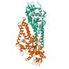
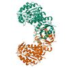
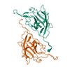
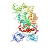
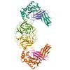
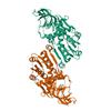
X-ray diffraction data for the Crystal Structure of 6,7-dimethyl-8-ribityllumazine synthase from Bordetella pertussis

First author:
Seattle Structural Genomics Center for Infectious Disease (SSGCID)
Uniprot: Q7VTN4
Resolution: 2.34 Å
R/Rfree: 0.21/0.26
Uniprot: Q7VTN4
Resolution: 2.34 Å
R/Rfree: 0.21/0.26
X-ray diffraction data for the Crystal Structure of 6,7-dimethyl-8-ribityllumazine synthase from Bordetella pertussis in complex with 6,7-dimethyl-8-(1'-D-ribityl) lumazine
First author:
Seattle Structural Genomics Center for Infectious Disease (SSGCID)
Uniprot: Q7VTN4
Resolution: 2.15 Å
R/Rfree: 0.22/0.26
Uniprot: Q7VTN4
Resolution: 2.15 Å
R/Rfree: 0.22/0.26
X-ray diffraction data for the Crystal structure of nucleoside-diphosphate kinase Cryptosporidium parvum (Apo, hexamer)
First author:
Seattle Structural Genomics Center for Infectious Disease (SSGCID) Seattle Structural Genomics Center for Infectious Disease
Uniprot: Q5CR64
Resolution: 1.48 Å
R/Rfree: 0.13/0.16
Uniprot: Q5CR64
Resolution: 1.48 Å
R/Rfree: 0.13/0.16
X-ray diffraction data for the Crystal structure of Phosphoribosylaminoimidazole carboxylase from Burkholderia xenovorans (ATP complex)

First author:
Seattle Structural Genomics Center for Infectious Disease (SSGCID) Seattle Structural Genomics Center for Infectious Disease
Uniprot: Q13UJ9
Resolution: 1.93 Å
R/Rfree: 0.17/0.20
Uniprot: Q13UJ9
Resolution: 1.93 Å
R/Rfree: 0.17/0.20
X-ray diffraction data for the Crystal structure of Phosphoribosylaminoimidazole carboxylase from Burkholderia xenovorans (AMP complex)

First author:
Seattle Structural Genomics Center for Infectious Disease (SSGCID) Seattle Structural Genomics Center for Infectious Disease
Uniprot: Q13UJ9
Resolution: 2.19 Å
R/Rfree: 0.17/0.20
Uniprot: Q13UJ9
Resolution: 2.19 Å
R/Rfree: 0.17/0.20
X-ray diffraction data for the Crystal structure of Glutamate--tRNA ligase (GltX) from Moraxella catarrhalis (Apo)
First author:
Seattle Structural Genomics Center for Infectious Disease (SSGCID) Seattle Structural Genomics Center for Infectious Disease
Uniprot: A0AB36DQE3
Resolution: 2.75 Å
R/Rfree: 0.22/0.26
Uniprot: A0AB36DQE3
Resolution: 2.75 Å
R/Rfree: 0.22/0.26
X-ray diffraction data for the Equine Serum Albumin with Copper(II)
X-ray diffraction data for the Crystal structure of calcium-dependent protein kinase 1 (CDPK1) from Cryptosporidium parvum in complex with inhibitor WIN-3-115
First author:
Seattle Structural Genomics Center for Infectious Disease (SSGCID) Seattle Structural Genomics Center for Infectious Disease
Uniprot: A3FQ16
Resolution: 2.94 Å
R/Rfree: 0.20/0.25
Uniprot: A3FQ16
Resolution: 2.94 Å
R/Rfree: 0.20/0.25
X-ray diffraction data for the Crystal structure of calcium-dependent protein kinase 1 (CDPK1) from Cryptosporidium parvum in complex with inhibitor BKI-1606
First author:
Seattle Structural Genomics Center for Infectious Disease (SSGCID) Seattle Structural Genomics Center for Infectious Disease
Uniprot: A3FQ16
Resolution: 2.75 Å
R/Rfree: 0.21/0.26
Uniprot: A3FQ16
Resolution: 2.75 Å
R/Rfree: 0.21/0.26
X-ray diffraction data for the Crystal structure of a calcium bound C2 domain containing protein from Trichomonas vaginalis
First author:
Seattle Structural Genomics Center for Infectious Disease (SSGCID)
Uniprot: A2EZR3
Resolution: 1.40 Å
R/Rfree: 0.14/0.17
Uniprot: A2EZR3
Resolution: 1.40 Å
R/Rfree: 0.14/0.17
X-ray diffraction data for the Crystal structure of a calcium bound C2 domain containing protein from Trichomonas vaginalis (P21 form)

First author:
Seattle Structural Genomics Center for Infectious Disease (SSGCID)
Uniprot: A2EZR3
Resolution: 1.51 Å
R/Rfree: 0.19/0.21
Uniprot: A2EZR3
Resolution: 1.51 Å
R/Rfree: 0.19/0.21
X-ray diffraction data for the Crystal structure of a calcium bound C2 domain containing protein from Trichomonas vaginalis (orthorhombic P form)
First author:
Seattle Structural Genomics Center for Infectious Disease (SSGCID)
Uniprot: A2EZR3
Resolution: 1.53 Å
R/Rfree: 0.23/0.26
Uniprot: A2EZR3
Resolution: 1.53 Å
R/Rfree: 0.23/0.26
X-ray diffraction data for the Crystal structure of a C2 domain containing protein from Trichomonas vaginalis
First author:
Seattle Structural Genomics Center for Infectious Disease (SSGCID)
Uniprot: A2EZR3
Resolution: 1.35 Å
R/Rfree: 0.15/0.17
Uniprot: A2EZR3
Resolution: 1.35 Å
R/Rfree: 0.15/0.17
X-ray diffraction data for the Crystal structure of a malonate bound C2 domain containing protein from Trichomonas vaginalis (P21 form)
First author:
Seattle Structural Genomics Center for Infectious Disease (SSGCID)
Uniprot: A2EZR3
Resolution: 1.30 Å
R/Rfree: 0.14/0.17
Uniprot: A2EZR3
Resolution: 1.30 Å
R/Rfree: 0.14/0.17
X-ray diffraction data for the Crystal structure of a calcium bound C2 domain containing protein from Trichomonas vaginalis (P61 form)
First author:
Seattle Structural Genomics Center for Infectious Disease (SSGCID)
Uniprot: A2EZR3
Resolution: 1.54 Å
R/Rfree: 0.17/0.20
Uniprot: A2EZR3
Resolution: 1.54 Å
R/Rfree: 0.17/0.20
X-ray diffraction data for the Crystal structure of a C2 domain containing protein from Trichomonas vaginalis in complex with pyrophosphate
First author:
Seattle Structural Genomics Center for Infectious Disease (SSGCID)
Uniprot: A2EZR3
Resolution: 1.70 Å
R/Rfree: 0.21/0.23
Uniprot: A2EZR3
Resolution: 1.70 Å
R/Rfree: 0.21/0.23
X-ray diffraction data for the Crystal structure of a Heat shock protein (DnaJ) from Brucella melitensis
First author:
Seattle Structural Genomics Center for Infectious Disease (SSGCID)
Uniprot: Q2YMC9
Resolution: 3.09 Å
R/Rfree: 0.24/0.29
Uniprot: Q2YMC9
Resolution: 3.09 Å
R/Rfree: 0.24/0.29
X-ray diffraction data for the Crystal structure of Phosphoglycerate mutase from Trichomonas vaginalis in complex with 3-phosphoglyceric acid
First author:
Seattle Structural Genomics Center for Infectious Disease (SSGCID) Seattle Structural Genomics Center for Infectious Disease
Uniprot: A2DUN8
Resolution: 2.10 Å
R/Rfree: 0.18/0.21
Uniprot: A2DUN8
Resolution: 2.10 Å
R/Rfree: 0.18/0.21
X-ray diffraction data for the Crystal structure of Glutamate-tRNA synthetase GluRS from Chlamydia pneumoniae (Orthorhombic C form)
First author:
Seattle Structural Genomics Center for Infectious Disease (SSGCID) Seattle Structural Genomics Center for Infectious Disease
Uniprot: Q9Z7Z3
Resolution: 2.75 Å
R/Rfree: 0.22/0.26
Uniprot: Q9Z7Z3
Resolution: 2.75 Å
R/Rfree: 0.22/0.26
X-ray diffraction data for the Crystal structure of Glutamate-tRNA synthetase GluRS from Chlamydia pneumoniae in complex with ATP
First author:
Seattle Structural Genomics Center for Infectious Disease (SSGCID) Seattle Structural Genomics Center for Infectious Disease
Uniprot: Q9Z7Z3
Resolution: 2.65 Å
R/Rfree: 0.21/0.26
Uniprot: Q9Z7Z3
Resolution: 2.65 Å
R/Rfree: 0.21/0.26
X-ray diffraction data for the Crystal Structure of CBS domain containing protein from Burkholderia phymatum
First author:
Seattle Structural Genomics Center for Infectious Disease (SSGCID)
Uniprot: B2JRV0
Resolution: 1.39 Å
R/Rfree: 0.16/0.19
Uniprot: B2JRV0
Resolution: 1.39 Å
R/Rfree: 0.16/0.19
X-ray diffraction data for the Crystal Structure of serine/threonine-protein kinase (AEK1) from Trypanosoma brucei
First author:
Seattle Structural Genomics Center for Infectious Disease (SSGCID)
Uniprot: Q582V7
Resolution: 2.05 Å
R/Rfree: 0.19/0.23
Uniprot: Q582V7
Resolution: 2.05 Å
R/Rfree: 0.19/0.23
X-ray diffraction data for the Crystal structure of a glyceraldehyde-3-phosphate dehydrogenase from Neisseria gonorrhoeae in complex with NAD (P1 form)

First author:
Seattle Structural Genomics Center for Infectious Disease (SSGCID)
Uniprot: B4RPP8
Resolution: 2.30 Å
R/Rfree: 0.19/0.23
Uniprot: B4RPP8
Resolution: 2.30 Å
R/Rfree: 0.19/0.23
X-ray diffraction data for the Crystal structure of a glyceraldehyde-3-phosphate dehydrogenase from Neisseria gonorrhoeae in complex with NAD and GLYCERALDEHYDE-3-PHOSPHATE

First author:
Seattle Structural Genomics Center for Infectious Disease (SSGCID) Seattle Structural Genomics Center for Infectious Disease
Uniprot: B4RPP8
Resolution: 1.91 Å
R/Rfree: 0.15/0.19
Uniprot: B4RPP8
Resolution: 1.91 Å
R/Rfree: 0.15/0.19
X-ray diffraction data for the Crystal structure of Formyl-coenzyme A transferase from Brucella melitensis in complex with succinate
First author:
Seattle Structural Genomics Center for Infectious Disease (SSGCID) Seattle Structural Genomics Center for Infectious Disease
Uniprot: Q8YDF2
Resolution: 2.88 Å
R/Rfree: 0.19/0.23
Uniprot: Q8YDF2
Resolution: 2.88 Å
R/Rfree: 0.19/0.23
X-ray diffraction data for the Crystal structure of Human Prostaglandin reductase 1 (PTGR1) in complex with NADP
First author:
Seattle Structural Genomics Center for Infectious Disease (SSGCID) Seattle Structural Genomics Center for Infectious Disease
Uniprot: Q14914
Resolution: 2.00 Å
R/Rfree: 0.17/0.21
Uniprot: Q14914
Resolution: 2.00 Å
R/Rfree: 0.17/0.21
X-ray diffraction data for the Crystal structure of Human Prostaglandin reductase 1 (PTGR1) in complex with NADP and Indomethacin
First author:
Seattle Structural Genomics Center for Infectious Disease (SSGCID) Seattle Structural Genomics Center for Infectious Disease
Uniprot: Q14914
Resolution: 2.10 Å
R/Rfree: 0.20/0.24
Uniprot: Q14914
Resolution: 2.10 Å
R/Rfree: 0.20/0.24
X-ray diffraction data for the Crystal structure of Human Prostaglandin reductase 1 (PTGR1) in complex with NADP and Indomethacin (Orthorhombic P form)
First author:
Seattle Structural Genomics Center for Infectious Disease (SSGCID) Seattle Structural Genomics Center for Infectious Disease
Uniprot: Q14914
Resolution: 2.30 Å
R/Rfree: 0.20/0.24
Uniprot: Q14914
Resolution: 2.30 Å
R/Rfree: 0.20/0.24
X-ray diffraction data for the Crystal structure of Glutamate-tRNA synthetase GluRS from Chlamydia pneumoniae in complex with O5'-(L-GLUTAMYL-SULFAMOYL)-ADENOSINE
First author:
Seattle Structural Genomics Center for Infectious Disease (SSGCID) Seattle Structural Genomics Center for Infectious Disease
Uniprot: Q9Z7Z3
Resolution: 2.80 Å
R/Rfree: 0.23/0.25
Uniprot: Q9Z7Z3
Resolution: 2.80 Å
R/Rfree: 0.23/0.25
X-ray diffraction data for the Structure of the Mus musclus Langerin carbohydrate recognition domain with depleted Calcium
First author:
M. Ruwolt
Resolution: 1.64 Å
R/Rfree: 0.15/0.21
Resolution: 1.64 Å
R/Rfree: 0.15/0.21
X-ray diffraction data for the Crystal structure of HLA-A0201 in complex with peptide LLWNGPMAV
X-ray diffraction data for the wt-1-9sl0

X-ray diffraction data for the Crystal structure of HLA-A0201 in complex with peptide SLLWNGPMAV
X-ray diffraction data for the Crystal structure of HLA-A0201 in complex with peptide LLWNGPMAVS
X-ray diffraction data for the Crystal structure of Cysteinyl-tRNA synthetase (CysRS) from Plasmodium falciparum (Hexagonal P form)
First author:
Seattle Structural Genomics Center for Infectious Disease (SSGCID)
Resolution: 2.87 Å
R/Rfree: 0.22/0.24
Resolution: 2.87 Å
R/Rfree: 0.22/0.24
X-ray diffraction data for the Crystal structure of Cysteinyl-tRNA synthetase (CysRS) from Plasmodium falciparum in complex with ATP (long soak)
First author:
Seattle Structural Genomics Center for Infectious Disease (SSGCID)
Uniprot: Q8IJP3
Resolution: 2.80 Å
R/Rfree: 0.20/0.22
Uniprot: Q8IJP3
Resolution: 2.80 Å
R/Rfree: 0.20/0.22
X-ray diffraction data for the Crystal structure of Cysteinyl-tRNA synthetase (CysRS) from Plasmodium falciparum in complex with AMP
First author:
Seattle Structural Genomics Center for Infectious Disease (SSGCID)
Uniprot: Q8IJP3
Resolution: 2.06 Å
R/Rfree: 0.18/0.20
Uniprot: Q8IJP3
Resolution: 2.06 Å
R/Rfree: 0.18/0.20
X-ray diffraction data for the Crystal structure of Cysteinyl-tRNA synthetase (CysRS) from Plasmodium falciparum in complex with GMP
First author:
Seattle Structural Genomics Center for Infectious Disease (SSGCID)
Uniprot: Q8IJP3
Resolution: 2.90 Å
R/Rfree: 0.19/0.22
Uniprot: Q8IJP3
Resolution: 2.90 Å
R/Rfree: 0.19/0.22
X-ray diffraction data for the Neutralizing monoclonal antibody Fab fragment bound to leptin

X-ray diffraction data for the Crystal structure of Cysteinyl-tRNA synthetase (CysRS) from Plasmodium falciparum in complex with O5'-(L-GLUTAMYL-SULFAMOYL)-ADENOSINE
First author:
Seattle Structural Genomics Center for Infectious Disease (SSGCID)
Uniprot: Q8IJP3
Resolution: 2.03 Å
R/Rfree: 0.18/0.20
Uniprot: Q8IJP3
Resolution: 2.03 Å
R/Rfree: 0.18/0.20
X-ray diffraction data for the Crystal structure of Cysteinyl-tRNA synthetase (CysRS) from Plasmodium falciparum in complex with Cysteine
First author:
Seattle Structural Genomics Center for Infectious Disease (SSGCID)
Uniprot: Q8IJP3
Resolution: 2.16 Å
R/Rfree: 0.19/0.22
Uniprot: Q8IJP3
Resolution: 2.16 Å
R/Rfree: 0.19/0.22
X-ray diffraction data for the Crystal structure of Cysteinyl-tRNA synthetase (CysRS) from Plasmodium falciparum in complex with ADP
First author:
Seattle Structural Genomics Center for Infectious Disease (SSGCID)
Uniprot: Q8IJP3
Resolution: 2.14 Å
R/Rfree: 0.18/0.20
Uniprot: Q8IJP3
Resolution: 2.14 Å
R/Rfree: 0.18/0.20
X-ray diffraction data for the Crystal structure of Cysteinyl-tRNA synthetase (CysRS) from Plasmodium falciparum in complex with AMP and Cysteine
First author:
Seattle Structural Genomics Center for Infectious Disease (SSGCID)
Uniprot: Q8IJP3
Resolution: 2.34 Å
R/Rfree: 0.19/0.22
Uniprot: Q8IJP3
Resolution: 2.34 Å
R/Rfree: 0.19/0.22
X-ray diffraction data for the Crystal structure of a Glyceraldehyde-3-phosphate dehydrogenase from Bordetella pertussis (monoclinic P form)
First author:
Seattle Structural Genomics Center for Infectious Disease (SSGCID) Seattle Structural Genomics Center for Infectious Disease
Uniprot: Q7VZB9
Resolution: 2.00 Å
R/Rfree: 0.17/0.20
Uniprot: Q7VZB9
Resolution: 2.00 Å
R/Rfree: 0.17/0.20
X-ray diffraction data for the Crystal Structure of Acetyl-CoA synthetase from Cryptococcus neoformans H99 in complex with inhibitor HGN-1310 (dd3-027)
First author:
Seattle Structural Genomics Center for Infectious Disease (SSGCID) Seattle Structural Genomics Center for Infectious Disease
Uniprot: J9VFT1
Resolution: 2.40 Å
R/Rfree: 0.18/0.22
Uniprot: J9VFT1
Resolution: 2.40 Å
R/Rfree: 0.18/0.22
X-ray diffraction data for the Rhombohedral crystalline form of human insulin complexed with m-cresol
X-ray diffraction data for the Crystal structure of Apo Cysteinyl-tRNA synthetase (CysRS) from Plasmodium falciparum (Orthrhombic P form)
First author:
Seattle Structural Genomics Center for Infectious Disease (SSGCID)
Uniprot: Q8IJP3
Resolution: 3.06 Å
R/Rfree: 0.23/0.29
Uniprot: Q8IJP3
Resolution: 3.06 Å
R/Rfree: 0.23/0.29
X-ray diffraction data for the Crystal structure of Cysteinyl-tRNA synthetase (CysRS) from Plasmodium falciparum in complex with 5'-Sulfamoyladenosine

First author:
Seattle Structural Genomics Center for Infectious Disease (SSGCID)
Uniprot: Q8IJP3
Resolution: 2.77 Å
R/Rfree: 0.19/0.23
Uniprot: Q8IJP3
Resolution: 2.77 Å
R/Rfree: 0.19/0.23
X-ray diffraction data for the Crystal structure of Cysteinyl-tRNA synthetase (CysRS) from Plasmodium falciparum in complex with cysteinyl-AMP
First author:
Seattle Structural Genomics Center for Infectious Disease (SSGCID)
Uniprot: Q8IJP3
Resolution: 2.22 Å
R/Rfree: 0.18/0.20
Uniprot: Q8IJP3
Resolution: 2.22 Å
R/Rfree: 0.18/0.20
X-ray diffraction data for the Structure of Saro_1862, a UPF0261 domain protein from Novosphingobium aromaticivorans with bound acetovanillone
X-ray diffraction data for the Crystal Structure of 6,7-dimethyl-8-ribityllumazine synthase from Bordetella pertussis in complex with 5-amino-6-(D-ribitylamino)uracil
First author:
Seattle Structural Genomics Center for Infectious Disease (SSGCID)
Uniprot: Q7VTN4
Resolution: 2.56 Å
R/Rfree: 0.20/0.25
Uniprot: Q7VTN4
Resolution: 2.56 Å
R/Rfree: 0.20/0.25
X-ray diffraction data for the Crystal structure of Glutamate--tRNA ligase (GltX) from Moraxella catarrhalis in complex with 5'-O-(N-Glutamyl)sulfamoyladeonosine
First author:
Seattle Structural Genomics Center for Infectious Disease (SSGCID) Seattle Structural Genomics Center for Infectious Disease
Uniprot: A0AB36DQE3
Resolution: 2.92 Å
R/Rfree: 0.21/0.25
Uniprot: A0AB36DQE3
Resolution: 2.92 Å
R/Rfree: 0.21/0.25
X-ray diffraction data for the Crystal structure of Prolyl-tRNA synthetase (ProRS, Proline--tRNA ligase) from Plasmodium falciparum in complex with inhibitor YNW69

First author:
Seattle Structural Genomics Center for Infectious Disease (SSGCID)
Uniprot: Q8I5R7
Resolution: 2.43 Å
R/Rfree: 0.20/0.23
Uniprot: Q8I5R7
Resolution: 2.43 Å
R/Rfree: 0.20/0.23
X-ray diffraction data for the Crystal structure of nucleoside-diphosphate kinase Cryptosporidium parvum (GMP complex)
First author:
Seattle Structural Genomics Center for Infectious Disease (SSGCID) Seattle Structural Genomics Center for Infectious Disease
Uniprot: Q5CR64
Resolution: 1.64 Å
R/Rfree: 0.15/0.18
Uniprot: Q5CR64
Resolution: 1.64 Å
R/Rfree: 0.15/0.18
X-ray diffraction data for the Crystal structure of phosphorylated C. merolae LAMMER-like dual specificity kinase (CmLIK) kinase domain in complex with adenosine
X-ray diffraction data for the Crystal structure of glutamate dehydrogenase from Babesia microti
First author:
Seattle Structural Genomics Center for Infectious Disease (SSGCID)
Uniprot: A0A0K3AUK4
Resolution: 2.18 Å
R/Rfree: 0.18/0.21
Uniprot: A0A0K3AUK4
Resolution: 2.18 Å
R/Rfree: 0.18/0.21
X-ray diffraction data for the Crystal structure of glutamate dehydrogenase from Babesia microti in complex with NADP
First author:
Seattle Structural Genomics Center for Infectious Disease (SSGCID)
Uniprot: A0A0K3AUK4
Resolution: 2.28 Å
R/Rfree: 0.19/0.22
Uniprot: A0A0K3AUK4
Resolution: 2.28 Å
R/Rfree: 0.19/0.22
X-ray diffraction data for the Crystal structure of HEI10
X-ray diffraction data for the Sal-Shy(DUF35) aldolase from Comamonas testosteroni
X-ray diffraction data for the GlfT2 from Nocardia brasiliensis
X-ray diffraction data for the Crystal Structure UTP--glucose-1-phosphate uridylyltransferase from Bordetella pertussis in complex with UTP
First author:
Seattle Structural Genomics Center for Infectious Disease (SSGCID)
Uniprot: Q7VTV0
Resolution: 1.82 Å
R/Rfree: 0.20/0.23
Uniprot: Q7VTV0
Resolution: 1.82 Å
R/Rfree: 0.20/0.23
X-ray diffraction data for the Crystal Structure UTP--glucose-1-phosphate uridylyltransferase from Bordetella pertussis in complex with URIDINE-5'-DIPHOSPHATE-GLUCOSE
First author:
Seattle Structural Genomics Center for Infectious Disease (SSGCID)
Uniprot: Q7VTV0
Resolution: 1.80 Å
R/Rfree: 0.17/0.20
Uniprot: Q7VTV0
Resolution: 1.80 Å
R/Rfree: 0.17/0.20
X-ray diffraction data for the Crystal Structure UTP--glucose-1-phosphate uridylyltransferase from Bordetella pertussis in complex with URIDINE-5'-DIPHOSPHATE-GLUCOSE (twinned lattice)
First author:
Seattle Structural Genomics Center for Infectious Disease (SSGCID)
Uniprot: Q7VTV0
Resolution: 1.68 Å
R/Rfree: 0.17/0.20
Uniprot: Q7VTV0
Resolution: 1.68 Å
R/Rfree: 0.17/0.20
X-ray diffraction data for the Crystal Structure UTP--glucose-1-phosphate uridylyltransferase from Bordetella pertussis (sulfate bound)
First author:
Seattle Structural Genomics Center for Infectious Disease (SSGCID)
Uniprot: Q7VTV0
Resolution: 1.95 Å
R/Rfree: 0.18/0.22
Uniprot: Q7VTV0
Resolution: 1.95 Å
R/Rfree: 0.18/0.22
X-ray diffraction data for the Crystal structure of a phage catechol 1,2-deoxygenase identified from a soil metagenomic survey.
First author:
Seattle Structural Genomics Center for Infectious Disease (SSGCID) Seattle Structural Genomics Center for Infectious Disease
Resolution: 1.65 Å
R/Rfree: 0.17/0.21
Resolution: 1.65 Å
R/Rfree: 0.17/0.21
X-ray diffraction data for the Crystal structure of DNA integrity scanning protein (DisA) from Mycobacterium tuberculosis in complex with cyclic di-AMP
First author:
Seattle Structural Genomics Center for Infectious Disease (SSGCID)
Uniprot: P9WNW5
Resolution: 2.89 Å
R/Rfree: 0.20/0.25
Uniprot: P9WNW5
Resolution: 2.89 Å
R/Rfree: 0.20/0.25
X-ray diffraction data for the Crystal structure of glyceraldehyde-3-phosphate dehydrogenase (GAPDH) from Neisseria gonorrhoeae in complex with NAD (Orthorhombic I form)

First author:
Seattle Structural Genomics Center for Infectious Disease (SSGCID)
Uniprot: B4RPP8
Resolution: 1.90 Å
R/Rfree: 0.18/0.21
Uniprot: B4RPP8
Resolution: 1.90 Å
R/Rfree: 0.18/0.21
X-ray diffraction data for the Crystal structure of Aeromonas salmonicida putative carbohydrate binding module and split domain
X-ray diffraction data for the Crystal structure of Glutamate-tRNA synthetase GluRS from Chlamydia pneumoniae
First author:
Seattle Structural Genomics Center for Infectious Disease (SSGCID)
Uniprot: Q9Z7Z3
Resolution: 2.08 Å
R/Rfree: 0.18/0.23
Uniprot: Q9Z7Z3
Resolution: 2.08 Å
R/Rfree: 0.18/0.23
X-ray diffraction data for the Crystal structure of Ornithine carbamoyltransferase from Burkholderia xenovorans
First author:
Seattle Structural Genomics Center for Infectious Disease (SSGCID)
Uniprot: Q13H08
Resolution: 2.55 Å
R/Rfree: 0.19/0.23
Uniprot: Q13H08
Resolution: 2.55 Å
R/Rfree: 0.19/0.23
X-ray diffraction data for the Crystal structure of Ornithine carbamoyltransferase from Burkholderia xenovorans in complex with phosphono carbamate
First author:
Seattle Structural Genomics Center for Infectious Disease (SSGCID)
Uniprot: Q13H08
Resolution: 2.67 Å
R/Rfree: 0.19/0.25
Uniprot: Q13H08
Resolution: 2.67 Å
R/Rfree: 0.19/0.25
X-ray diffraction data for the Crystal structure of DNA integrity scanning protein DisA from Mycobacterium tuberculosis in complex with cyclic di-AMP and bromide
First author:
Seattle Structural Genomics Center for Infectious Disease (SSGCID)
Uniprot: P9WNW5
Resolution: 3.03 Å
R/Rfree: 0.23/0.27
Uniprot: P9WNW5
Resolution: 3.03 Å
R/Rfree: 0.23/0.27
X-ray diffraction data for the Crystal structure of a SnoaL-like domain-containing protein from Mycobacterium ulcerans (Orthorhombic C form)
First author:
Seattle Structural Genomics Center for Infectious Disease (SSGCID) Seattle Structural Genomics Center for Infectious Disease
Uniprot: A0PR67
Resolution: 1.36 Å
R/Rfree: 0.16/0.18
Uniprot: A0PR67
Resolution: 1.36 Å
R/Rfree: 0.16/0.18
X-ray diffraction data for the 293K human S-adenosylmethionine decarboxylase
X-ray diffraction data for the 100K human S-adenosylmethionine decarboxylase
X-ray diffraction data for the Crystal structure of D-aspartate oxidase from Cryptococcus humicola UJ1.

X-ray diffraction data for the A chimera FadR transcription factor containing wHTH of Rv0494 and the part other than wHTH of Escherichia coli FadR
X-ray diffraction data for the Klebsiella pneumoniae maltohexaose-producing alpha-amylase
X-ray diffraction data for the Klebsiella pneumoniae maltohexaose-producing alpha-amylase in complex with acarbose
X-ray diffraction data for the Klebsiella pneumoniae maltohexaose-producing alpha-amylase, terbium derivative
X-ray diffraction data for the 273K human S-adenosylmethionine decarboxylase
X-ray diffraction data for the Crystal structure of human astrovirus 1 capsid spike bound to human neonatal Fc receptor
X-ray diffraction data for the Bacillus licheniformis nitroreductase
X-ray diffraction data for the Crystal structure of M. smegmatis GMP reductase in complex with IMP.
X-ray diffraction data for the Crystal structure of M. smegmatis GMP reductase in complex with GMP and ATP.
X-ray diffraction data for the Crystal structure of M. smegmatis GMP reductase in complex with GMP and GTP.
X-ray diffraction data for the Crystal structure of M. smegmatis GMP reductase in complex with IMP and ATP.
X-ray diffraction data for the Crystal structure of Apo Human Prostaglandin reductase 1 (PTGR1) (Monoclinic P form)
First author:
Seattle Structural Genomics Center for Infectious Disease (SSGCID) Seattle Structural Genomics Center for Infectious Disease
Uniprot: Q14914
Resolution: 2.10 Å
R/Rfree: 0.21/0.24
Uniprot: Q14914
Resolution: 2.10 Å
R/Rfree: 0.21/0.24
X-ray diffraction data for the Crystal structure of Apo Human Prostaglandin reductase 1 (PTGR1) (Orthorhombic C form)
First author:
Seattle Structural Genomics Center for Infectious Disease (SSGCID) Seattle Structural Genomics Center for Infectious Disease
Uniprot: Q14914
Resolution: 2.35 Å
R/Rfree: 0.20/0.25
Uniprot: Q14914
Resolution: 2.35 Å
R/Rfree: 0.20/0.25
X-ray diffraction data for the Crystal structure of Human Prostaglandin reductase 1 (PTGR1) in complex with methotrexate (P1 form)
First author:
Seattle Structural Genomics Center for Infectious Disease (SSGCID) Seattle Structural Genomics Center for Infectious Disease
Uniprot: Q14914
Resolution: 2.00 Å
R/Rfree: 0.21/0.25
Uniprot: Q14914
Resolution: 2.00 Å
R/Rfree: 0.21/0.25
X-ray diffraction data for the Crystal structure of Human Prostaglandin reductase 1 (PTGR1) in complex with methotrexate (Monoclinic P form)
First author:
Seattle Structural Genomics Center for Infectious Disease (SSGCID) Seattle Structural Genomics Center for Infectious Disease
Uniprot: Q14914
Resolution: 1.95 Å
R/Rfree: 0.18/0.22
Uniprot: Q14914
Resolution: 1.95 Å
R/Rfree: 0.18/0.22
X-ray diffraction data for the Crystal structure of Human Prostaglandin reductase 1 (PTGR1) in complex with methotrexate (Orthorhombic C form)
First author:
Seattle Structural Genomics Center for Infectious Disease (SSGCID) Seattle Structural Genomics Center for Infectious Disease
Uniprot: Q14914
Resolution: 1.75 Å
R/Rfree: 0.17/0.19
Uniprot: Q14914
Resolution: 1.75 Å
R/Rfree: 0.17/0.19
X-ray diffraction data for the Crystal structure of Uracil-DNA glycosylase from Burkholderia thailandensis
First author:
Seattle Structural Genomics Center for Infectious Disease (SSGCID) Seattle Structural Genomics Center for Infectious Disease
Uniprot: Q2SUH8
Resolution: 1.70 Å
R/Rfree: 0.16/0.20
Uniprot: Q2SUH8
Resolution: 1.70 Å
R/Rfree: 0.16/0.20
X-ray diffraction data for the Crystal structure of SpMETTL16 kinase associated 1 domain in complex with U6 snRNA internal stem loop
X-ray diffraction data for the Crystal structure of Capsular polysaccharide biosynthesis protein from Bordetella pertussis in complex with NAD and uridine-diphosphate-n-acetylgalactosamine
First author:
Seattle Structural Genomics Center for Infectious Disease (SSGCID) Seattle Structural Genomics Center for Infectious Disease
Uniprot: Q7TTK0
Resolution: 2.40 Å
R/Rfree: 0.19/0.25
Uniprot: Q7TTK0
Resolution: 2.40 Å
R/Rfree: 0.19/0.25
X-ray diffraction data for the Crystal structure of a SnoaL-like domain-containing protein from Mycobacterium ulcerans

First author:
Seattle Structural Genomics Center for Infectious Disease (SSGCID) Seattle Structural Genomics Center for Infectious Disease
Uniprot: A0PR67
Resolution: 1.37 Å
R/Rfree: 0.15/0.19
Uniprot: A0PR67
Resolution: 1.37 Å
R/Rfree: 0.15/0.19
X-ray diffraction data for the Crystal structure of nucleoside-diphosphate kinase Cryptosporidium parvum in complex with ATP
First author:
Seattle Structural Genomics Center for Infectious Disease (SSGCID) Seattle Structural Genomics Center for Infectious Disease
Uniprot: Q5CR64
Resolution: 1.42 Å
R/Rfree: 0.13/0.16
Uniprot: Q5CR64
Resolution: 1.42 Å
R/Rfree: 0.13/0.16
X-ray diffraction data for the Crystal structure of nucleoside-diphosphate kinase Cryptosporidium parvum in complex with AMP
First author:
Seattle Structural Genomics Center for Infectious Disease (SSGCID) Seattle Structural Genomics Center for Infectious Disease
Uniprot: Q5CR64
Resolution: 1.45 Å
R/Rfree: 0.12/0.16
Uniprot: Q5CR64
Resolution: 1.45 Å
R/Rfree: 0.12/0.16
X-ray diffraction data for the Crystal structure of Glyceraldehyde-3-phosphate dehydrogenase from Bordetella pertussis (NAD bound)
First author:
Seattle Structural Genomics Center for Infectious Disease (SSGCID) Seattle Structural Genomics Center for Infectious Disease
Uniprot: Q7VZB9
Resolution: 2.15 Å
R/Rfree: 0.19/0.23
Uniprot: Q7VZB9
Resolution: 2.15 Å
R/Rfree: 0.19/0.23