6929 results
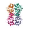
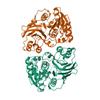
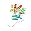
X-ray diffraction data for the Dihydroneopterin aldolase/dihydroneopterin triphosphate 2'-epimerase from Yersinia pestis.

First author:
J. Osipiuk
Gene name: folB
Resolution: 2.15 Å
R/Rfree: 0.22/0.26
Gene name: folB
Resolution: 2.15 Å
R/Rfree: 0.22/0.26
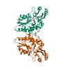
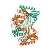
X-ray diffraction data for the 1.85 Angstrom resolution crystal structure of an ABC transporter from Clostridium perfringens ATCC 13124
First author:
A.S. Halavaty
Resolution: 1.85 Å
R/Rfree: 0.16/0.19
Resolution: 1.85 Å
R/Rfree: 0.16/0.19
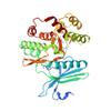
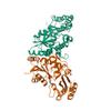
X-ray diffraction data for the 2.25 Angstrom resolution crystal structure of UDP-N-acetylmuramate--L-alanine ligase (murC) from Yersinia pestis CO92 in complex with AMP
First author:
A.S. Halavaty
Gene name: murC
Resolution: 2.25 Å
R/Rfree: 0.18/0.22
Gene name: murC
Resolution: 2.25 Å
R/Rfree: 0.18/0.22
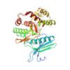
X-ray diffraction data for the Crystal structure of bifunctional folylpolyglutamate synthase/dihydrofolate synthase with Mn, AMPPNP and L-Glutamate bound
First author:
B. Nocek
Gene name: folC
Resolution: 2.00 Å
R/Rfree: 0.17/0.20
Gene name: folC
Resolution: 2.00 Å
R/Rfree: 0.17/0.20
X-ray diffraction data for the 2.0 Angstrom Crystal Structure of Ligand Binding Component of ABC-type Import System from Staphylococcus aureus with Zinc bound
First author:
G. Minasov
Resolution: 2.01 Å
R/Rfree: 0.16/0.21
Resolution: 2.01 Å
R/Rfree: 0.16/0.21
X-ray diffraction data for the Crystal structure of the 3-oxoacyl-acyl carrier protein reductase, FabG, from Staphylococcus aureus
First author:
S.M. Anderson
Gene name: fabG
Resolution: 1.90 Å
R/Rfree: 0.16/0.19
Gene name: fabG
Resolution: 1.90 Å
R/Rfree: 0.16/0.19
X-ray diffraction data for the 2.5 Angstrom Resolution Crystal Structure of 3-Dehydroquinate Synthase (aroB) from Vibrio cholerae
First author:
G. Minasov
Gene name: aroB
Resolution: 2.50 Å
R/Rfree: 0.18/0.22
Gene name: aroB
Resolution: 2.50 Å
R/Rfree: 0.18/0.22
X-ray diffraction data for the 2.4 Angstrom Crystal Structure of Dihydroorotase (pyrC) from Campylobacter jejuni.
First author:
G. Minasov
Gene name: pyrC
Resolution: 2.40 Å
R/Rfree: 0.19/0.25
Gene name: pyrC
Resolution: 2.40 Å
R/Rfree: 0.19/0.25
X-ray diffraction data for the Crystal structure of the aminoglycoside phosphotransferase APH(3')-Ia, with substrate kanamycin and small molecule inhibitor 1-NM-PP1
First author:
P.J. Stogios
Gene name: aphA1
Resolution: 1.88 Å
R/Rfree: 0.17/0.22
Gene name: aphA1
Resolution: 1.88 Å
R/Rfree: 0.17/0.22
X-ray diffraction data for the Crystal Structure of Superantigen-like Protein, Exotoxin SACOL0473 from Staphylococcus aureus subsp. aureus COL

First author:
E.V. Filippova
Resolution: 2.30 Å
R/Rfree: 0.19/0.25
Resolution: 2.30 Å
R/Rfree: 0.19/0.25
X-ray diffraction data for the Crystal structure of menaquinone-specific isochorismate synthase from Yersinia pestis CO92
First author:
B. Nocek
Gene name: menF
Resolution: 2.28 Å
R/Rfree: 0.19/0.25
Gene name: menF
Resolution: 2.28 Å
R/Rfree: 0.19/0.25
X-ray diffraction data for the 2.23 Angstrom resolution crystal structure of UDP-N-acetylglucosamine 1-carboxyvinyltransferase (murA) from Listeria monocytogenes EGD-e
First author:
A.S. Halavaty
Gene name: murA
Resolution: 2.23 Å
R/Rfree: 0.20/0.24
Gene name: murA
Resolution: 2.23 Å
R/Rfree: 0.20/0.24
X-ray diffraction data for the 1.95 Angstrom Resolution Crystal Structure of 3-deoxy-D-manno-octulosonate 8-phosphate phosphatase from Yersinia pestis
First author:
G. Minasov
Resolution: 1.95 Å
R/Rfree: 0.16/0.20
Resolution: 1.95 Å
R/Rfree: 0.16/0.20
X-ray diffraction data for the 2.05 Angstrom Crystal Structure of Ribulose-phosphate 3-epimerase from Toxoplasma gondii.
First author:
G. Minasov
Resolution: 2.05 Å
R/Rfree: 0.15/0.19
Resolution: 2.05 Å
R/Rfree: 0.15/0.19
X-ray diffraction data for the 2.1 Angstrom Resolution Crystal Structure of Metallo-beta-lactamase from Staphylococcus aureus subsp. aureus COL
First author:
G. Minasov
Resolution: 2.10 Å
R/Rfree: 0.16/0.20
Resolution: 2.10 Å
R/Rfree: 0.16/0.20
X-ray diffraction data for the Crystal Structure of Pantoate-beta-alanine Ligase from Francisella tularensis Complexed with Beta-gamma ATP and Beta-alanine
First author:
N. Maltseva
Gene name: panC
Resolution: 2.60 Å
R/Rfree: 0.17/0.25
Gene name: panC
Resolution: 2.60 Å
R/Rfree: 0.17/0.25
X-ray diffraction data for the Crystal structure of the aminoglycoside phosphotransferase APH(3')-Ia, with substrate kanamycin and small molecule inhibitor tyrphostin AG1478
First author:
P.J. Stogios
Gene name: aphA1
Resolution: 2.71 Å
R/Rfree: 0.18/0.24
Gene name: aphA1
Resolution: 2.71 Å
R/Rfree: 0.18/0.24
X-ray diffraction data for the Crystal structure of the aminoglycoside phosphotransferase APH(3')-Ia, with substrate kanamycin and small molecule inhibitor anthrapyrazolone SP600125
First author:
P.J. Stogios
Gene name: aphA1
Resolution: 2.37 Å
R/Rfree: 0.16/0.21
Gene name: aphA1
Resolution: 2.37 Å
R/Rfree: 0.16/0.21
X-ray diffraction data for the Crystal structure of the S112A mutant of mycrocine immunity protein (MccF) with AMP
First author:
B. Nocek
Resolution: 1.75 Å
R/Rfree: 0.19/0.20
Resolution: 1.75 Å
R/Rfree: 0.19/0.20
X-ray diffraction data for the Crystal structure of aminoglycoside 4'-O-adenylyltransferase ANT(4')-IIb, tobramycin-bound
First author:
P.J. Stogios
Gene name: ant4'-IIb
Resolution: 2.15 Å
R/Rfree: 0.17/0.22
Gene name: ant4'-IIb
Resolution: 2.15 Å
R/Rfree: 0.17/0.22
X-ray diffraction data for the Crystal structure of aminoglycoside 4'-O-adenylyltransferase ANT(4')-IIb, apo

First author:
P.J. Stogios
Gene name: ant4'-IIb
Resolution: 1.60 Å
R/Rfree: 0.17/0.19
Gene name: ant4'-IIb
Resolution: 1.60 Å
R/Rfree: 0.17/0.19
X-ray diffraction data for the Pantoate-beta-alanine ligase from Yersinia pestis
First author:
J. Osipiuk
Gene name: panC
Resolution: 1.83 Å
R/Rfree: 0.17/0.21
Gene name: panC
Resolution: 1.83 Å
R/Rfree: 0.17/0.21
X-ray diffraction data for the 1.85 Angstrom Resolution Crystal Structure of Transaldolase B (talA) from Francisella tularensis.
First author:
G. Minasov
Gene name: talA
Resolution: 1.85 Å
R/Rfree: 0.15/0.19
Gene name: talA
Resolution: 1.85 Å
R/Rfree: 0.15/0.19
X-ray diffraction data for the Crystal structure of the aminoglycoside phosphotransferase APH(3')-Ia, with substrate kanamycin and small molecule inhibitor pyrazolopyrimidine PP1
First author:
P.J. Stogios
Gene name: aphA1
Resolution: 1.89 Å
R/Rfree: 0.15/0.20
Gene name: aphA1
Resolution: 1.89 Å
R/Rfree: 0.15/0.20
X-ray diffraction data for the Crystal structure of the aminoglycoside phosphotransferase APH(3')-Ia, with substrate kanamycin and small molecule inhibitor pyrazolopyrimidine PP2
First author:
P.J. Stogios
Gene name: aphA1
Resolution: 1.98 Å
R/Rfree: 0.16/0.22
Gene name: aphA1
Resolution: 1.98 Å
R/Rfree: 0.16/0.22