6914 results
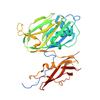
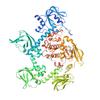
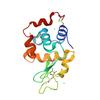
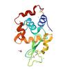
X-ray diffraction data for the D-GlcNAc-bound structure of Vibrio vulnificus putative carbohydrate binding module and split domain
First author:
B.P. Soares
Resolution: 2.28 Å
R/Rfree: 0.19/0.22
Resolution: 2.28 Å
R/Rfree: 0.19/0.22
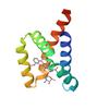
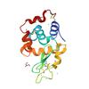
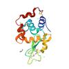
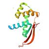
X-ray diffraction data for the Structure of de novo designed doxorubicin binding protein with doxorubicin bound
First author:
M. Horst
Resolution: 1.56 Å
R/Rfree: 0.22/0.26
Resolution: 1.56 Å
R/Rfree: 0.22/0.26
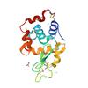
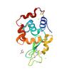
X-ray diffraction data for the Crystal Structure of cGMP-dependent protein kinase from Plasmodium vivax in complex with inhibitor RUBP-60
First author:
Seattle Structural Genomics Center for Infectious Disease (SSGCID) Seattle Structural Genomics Center for Infectious Disease
Uniprot: A5K0N4
Resolution: 2.90 Å
R/Rfree: 0.22/0.27
Uniprot: A5K0N4
Resolution: 2.90 Å
R/Rfree: 0.22/0.27
X-ray diffraction data for the Crystal structure of hen egg white lysozyme at 100 Kelvin with PEG 6000
X-ray diffraction data for the Crystal structure of hen egg white lysozyme at 100 Kelvin with PEG 400 (Triplicate)

X-ray diffraction data for the Crystal structure of hen egg white lysozyme at 100 Kelvin with PEG 6000 (Duplicate)
X-ray diffraction data for the Crystal structure of hen egg white lysozyme at 100 Kelvin with PEG 400 (Duplicate)
X-ray diffraction data for the Crystal structure of hen egg white lysozyme at 100 Kelvin with PEG 6000 (Triplicate)

X-ray diffraction data for the Crystal structure of hen egg white lysozyme at 100 Kelvin with Silicone Oil (Triplicate)
X-ray diffraction data for the Crystal structure of hen egg white lysozyme at 100 Kelvin with Vaseline (Duplicate)
X-ray diffraction data for the Crystal structure of hen egg white lysozyme at 100 Kelvin with Vaseline
X-ray diffraction data for the Crystal structure of hen egg white lysozyme at 100 Kelvin with Silicone Oil

X-ray diffraction data for the Crystal structure of hen egg white lysozyme at 100 Kelvin with Silicone Oil (Duplicate)

X-ray diffraction data for the Crystal structure of hen egg white lysozyme at 300 Kelvin with Apiezon N (Triplicate)

X-ray diffraction data for the Crystal structure of hen egg white lysozyme at 100 Kelvin with Vaseline (Triplicate)
X-ray diffraction data for the Crystal structure of hen egg white lysozyme at 300 Kelvin with Vaseline
X-ray diffraction data for the Crystal structure of hen egg white lysozyme at 300 Kelvin with Apiezon N
X-ray diffraction data for the Crystal structure of hen egg white lysozyme at 300 Kelvin with Vaseline (Duplicate)
X-ray diffraction data for the Crystal structure of hen egg white lysozyme at 300 Kelvin with Vaseline (Triplicate)

X-ray diffraction data for the Crystal structure of hen egg white lysozyme at 300 Kelvin with Apiezon T (Triplicate)

X-ray diffraction data for the Crystal structure of hen egg white lysozyme at 300 Kelvin with Apiezon T (Duplicate)

X-ray diffraction data for the Crystal structure of hen egg white lysozyme at 300 Kelvin with Apiezon T
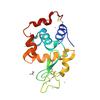
X-ray diffraction data for the Crystal structure of a large ribosomal subunit protein bL17 from Bordetella pertussis
First author:
Seattle Structural Genomics Center for Infectious Disease (SSGCID)
Uniprot: Q7VTA6
Resolution: 1.65 Å
R/Rfree: 0.19/0.22
Uniprot: Q7VTA6
Resolution: 1.65 Å
R/Rfree: 0.19/0.22
X-ray diffraction data for the psaea_00612_a_b3-36lw
First author:
Diffraction Data Upload SSGCID
X-ray diffraction data for the The dose series study of NcAA9D: wedge 20-38
First author:
Sam Miller