Diffraction project datasets IDP01801_3msz
- Method: Molecular Replacement
- Resolution: 2.053 Å
- Space group: P 32 2 1
PDB website for 3MSZ
PDB validation report for 3MSZ
doi:10.18430/m33msz
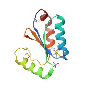
Project details
| Title | Crystal Structure of Glutaredoxin 1 from Francisella tularensis Complexed with Cacodylate |
| Authors | Maltseva, N., Kim, Y., Kwon, K., Anderson, W.F., Joachimiak, A., Center for Structural Genomics of Infectious Diseases (CSGID) |
| R / Rfree | 0.17 / 0.22 |
| Unit cell edges [Å] | 49.14 x 49.14 x 140.20 |
| Unit cell angles [°] | 90.0, 90.0, 120.0 |
Dataset ia6su-peak.####.img details
| Number of frames | 136 (1 - 136) |
| Distance [mm] | 310.0 |
| Oscillation width [°] | 1.00 |
| Omega [°] | -120.0 |
| Kappa / Chi [°] | 0.0002 |
| Phi [°] | -0.0108 |
| Wavelength [Å] | 0.97929 |
| Experiment Date | 2010-03-27 |
| Equipment | 19-ID at APS (Advanced Photon Source) |