Diffraction project datasets IDP04345_3pns
- Method: Molecular Replacement
- Resolution: 2.002 Å
- Space group: P 21 21 21
PDB website for 3PNS
PDB validation report for 3PNS
doi:10.18430/m33pns
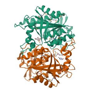
Project details
| Title | Crystal Structure of Uridine Phosphorylase Complexed with Uracil from Vibrio cholerae O1 biovar El Tor |
| Authors | Maltseva, N., Kim, Y., Hasseman, J., Anderson, W.F., Joachimiak, A., Center for Structural Genomics of Infectious Diseases (CSGID) |
| R / Rfree | 0.17 / 0.23 |
| Unit cell edges [Å] | 103.28 x 174.46 x 180.02 |
| Unit cell angles [°] | 90.0, 90.0, 90.0 |
Dataset urac-ps2e5g-peak.####.img details
| Number of frames | 480 (1 - 480) |
| Distance [mm] | 300.0 |
| Oscillation width [°] | 0.50 |
| Omega [°] | -120.0 |
| Kappa / Chi [°] | 0.0003 |
| Phi [°] | -0.0108 |
| Wavelength [Å] | 0.97921 |
| Experiment Date | 2010-08-09 |
| Equipment | 19-ID at APS (Advanced Photon Source) |