Diffraction project datasets MytuD00783aA1_PLP-aa_6uld
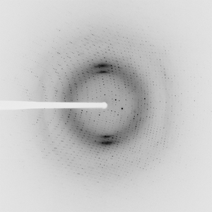
- Resolution: 1.5 Å
- Space group: P 1 21 1
PDB website for 6ULD
PDB validation report for 6ULD
doi:10.18430/m36uld
Project details
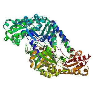
| Title | Crystal structure of serine hydroxymethyltransferase from Mycobacterium tuberculosis with bound PLP forming a Schiff base with substrate Serine in one monomer and PLP forming a Schiff base with product Glycine in the other monomer |
| Authors | Seattle Structural Genomics Center for Infectious Disease (SSGCID) |
| R / Rfree | 0.13 / 0.16 |
| Unit cell edges [Å] | 63.65 x 60.01 x 101.66 |
| Unit cell angles [°] | 90.0, 93.0, 90.0 |
Dataset vjp0-1.### details
| Number of frames | 200 (1 - 200) |
| Distance [mm] | 200.0 |
| Oscillation width [°] | 1.00 |
| Phi [°] | 100.0 |
| Wavelength [Å] | 0.97872 |
| Experiment Date | 2019-08-08 |
| Equipment | 21-ID-F at APS (Advanced Photon Source) |