Diffraction project datasets PX15607B_3hwu
Project details
| Title |
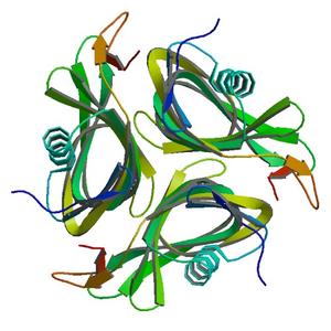
Crystal structure of Putative DNA-binding protein (YP_299413.1) from Ralstonia eutrophA JMP134 at 1.30 A resolution |
| Authors |
Joint Center for Structural Genomics (JCSG) |
| Bioentity |
None |
| R / Rfree |
0.12 / 0.14 |
| Unit cell edges [Å] |
53.90 x
53.90 x
111.10
|
| Unit cell angles [°] |
90.0,
90.0,
120.0
|
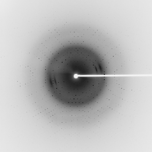
Dataset 114340_1_E1_###.mccd details
| Number of frames |
90 (1 - 90) |
| Distance [mm] |
200.0 |
| Oscillation width [°] |
1.00 |
| Phi [°] |
219.0 |
| Wavelength [Å] |
0.97929 |
| Experiment Date |
2009-03-19 |
| Equipment |
BL9-2
at SSRL (Stanford Synchrotron Radiation Laboratory)
|
Dataset 114340_1_E2_###.mccd details
| Number of frames |
90 (1 - 90) |
| Distance [mm] |
200.0 |
| Oscillation width [°] |
1.00 |
| Phi [°] |
219.0 |
| Wavelength [Å] |
0.91162 |
| Experiment Date |
2009-03-19 |
| Equipment |
BL9-2
at SSRL (Stanford Synchrotron Radiation Laboratory)
|
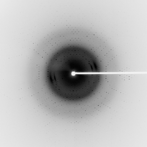
Dataset 114340_2_###.mccd details
| Number of frames |
90 (1 - 90) |
| Distance [mm] |
200.0 |
| Oscillation width [°] |
1.00 |
| Phi [°] |
219.0 |
| Wavelength [Å] |
0.97918 |
| Experiment Date |
2009-03-19 |
| Equipment |
BL9-2
at SSRL (Stanford Synchrotron Radiation Laboratory)
|