114 results
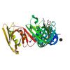
X-ray diffraction data for the Crystal structure of a DNA polymerase III subunit beta DnaN sliding clamp from Rickettsia typhi str. Wilmington
First author:
T.E. Edwards
Resolution: 2.00 Å
R/Rfree: 0.17/0.22
Resolution: 2.00 Å
R/Rfree: 0.17/0.22
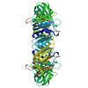
X-ray diffraction data for the Crystal structure of a DNA polymerase III subunit beta DnaN sliding clamp from Mycobacterium marinum
First author:
T.E. Edwards
Resolution: 2.45 Å
R/Rfree: 0.19/0.23
Resolution: 2.45 Å
R/Rfree: 0.19/0.23
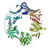
X-ray diffraction data for the Crystal structure of a DNA polymerase III subunit beta DnaN sliding clamp from Bartonella birtlesii LL-WM9
First author:
T.E. Edwards
Resolution: 2.45 Å
R/Rfree: 0.22/0.26
Resolution: 2.45 Å
R/Rfree: 0.22/0.26
First author:
K. Liu
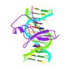
Resolution: 2.01 Å
R/Rfree: 0.21/0.25
Resolution: 2.01 Å
R/Rfree: 0.21/0.25
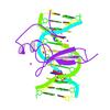
First author:
K. Liu
Resolution: 1.90 Å
R/Rfree: 0.22/0.25
Resolution: 1.90 Å
R/Rfree: 0.22/0.25
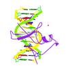
First author:
K. Liu
Resolution: 2.65 Å
R/Rfree: 0.21/0.24
Resolution: 2.65 Å
R/Rfree: 0.21/0.24
X-ray diffraction data for the Ultra-high resolution structure of d(CGCGCG)2 Z-DNA
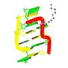
First author:
K. Brzezinski
Resolution: 0.55 Å
R/Rfree: 0.08/0.09
Resolution: 0.55 Å
R/Rfree: 0.08/0.09
X-ray diffraction data for the Crystal Structure of dnaN DNA polymerase III beta subunit from Stenotrophomonas maltophilia K279a
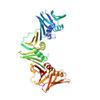
First author:
Seattle Structural Genomics Center for Infectious Disease (SSGCID)
Resolution: 2.15 Å
R/Rfree: 0.17/0.22
Resolution: 2.15 Å
R/Rfree: 0.17/0.22
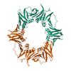
X-ray diffraction data for the Crystal structure of a DnaN sliding clamp (DNA polymerase III subunit beta) from Bartonella birtlesii bound to griselimycin
First author:
Seattle Structural Genomics Center for Infectious Disease (SSGCID)
Resolution: 1.75 Å
R/Rfree: 0.18/0.21
Resolution: 1.75 Å
R/Rfree: 0.18/0.21
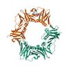
X-ray diffraction data for the Crystal structure of a DnaN sliding clamp (DNA polymerase III subunit beta) from Rickettsia rickettsii bound to griselimycin
First author:
Seattle Structural Genomics Center for Infectious Disease (SSGCID)
Resolution: 1.85 Å
R/Rfree: 0.17/0.21
Resolution: 1.85 Å
R/Rfree: 0.17/0.21
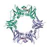
X-ray diffraction data for the Crystal structure of a DnaN sliding clamp (DNA polymerase III subunit beta) from Pseudomonas aeruginosa bound to griselimycin
First author:
Seattle Structural Genomics Center for Infectious Disease (SSGCID)
Resolution: 3.05 Å
R/Rfree: 0.19/0.22
Resolution: 3.05 Å
R/Rfree: 0.19/0.22
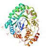
X-ray diffraction data for the Structure of a putative reductase from Yersinia pestis
X-ray diffraction data for the Mechanism of protease dependent DPC repair

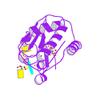
First author:
F. Li
Resolution: 1.55 Å
R/Rfree: 0.16/0.19
Resolution: 1.55 Å
R/Rfree: 0.16/0.19
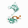
X-ray diffraction data for the C-Myc DNA binding protein complex
First author:
P. Aggarwal
Resolution: 2.57 Å
R/Rfree: 0.21/0.24
Resolution: 2.57 Å
R/Rfree: 0.21/0.24
X-ray diffraction data for the Crystal structure of a DnaN sliding clamp DNA polymerase III subunit beta from Rickettsia bellii RML369-C
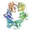
First author:
Seattle Structural Genomics Center for Infectious Disease (SSGCID) Seattle Structural Genomics Center for Infectious Disease
Resolution: 2.35 Å
R/Rfree: 0.19/0.24
Resolution: 2.35 Å
R/Rfree: 0.19/0.24
X-ray diffraction data for the HhaI endonuclease in Complex with Iodine-Labelled DNA

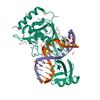
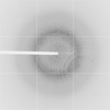
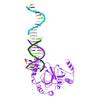
First author:
S. Qin
Resolution: 1.80 Å
R/Rfree: 0.20/0.23
Resolution: 1.80 Å
R/Rfree: 0.20/0.23
X-ray diffraction data for the MeCP2 MBD in complex with DNA

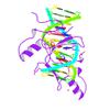
First author:
M. Lei
Resolution: 1.80 Å
R/Rfree: 0.23/0.26
Resolution: 1.80 Å
R/Rfree: 0.23/0.26

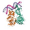
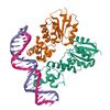
X-ray diffraction data for the Full-length dimer of DNA-Damage Response Protein C from Deinococcus radiodurans - Crystal form xMJ7124
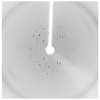
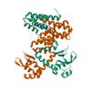
First author:
R. Szabla
Resolution: 4.28 Å
R/Rfree: 0.25/0.35
Resolution: 4.28 Å
R/Rfree: 0.25/0.35
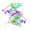
First author:
M. Lei
Resolution: 2.30 Å
R/Rfree: 0.23/0.27
Resolution: 2.30 Å
R/Rfree: 0.23/0.27
X-ray diffraction data for the Complex of MBD1-MBD and methylated DNA
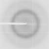
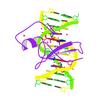
First author:
K. Liu
Resolution: 2.25 Å
R/Rfree: 0.24/0.27
Resolution: 2.25 Å
R/Rfree: 0.24/0.27
X-ray diffraction data for the Structure of apurinic/apyrimidinic DNA lyase TK0353 from Thermococcus kodakarensis (Iodide crystal form)

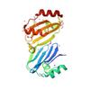
First author:
B.E. Eckenroth
Resolution: 2.20 Å
R/Rfree: 0.23/0.26
Resolution: 2.20 Å
R/Rfree: 0.23/0.26
X-ray diffraction data for the Structure of apurinic/apyrimidinic DNA lyase TK0353 from Thermococcus kodakarensis (Selenomethionine)

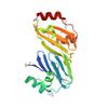
First author:
B.E. Eckenroth
Resolution: 1.98 Å
R/Rfree: 0.20/0.23
Resolution: 1.98 Å
R/Rfree: 0.20/0.23