402 results
X-ray diffraction data for the Crystal structure of a malate synthase G from Mycobacterium marinum bound to acetyl CoA
First author:
T.E. Edwards
Resolution: 1.60 Å
R/Rfree: 0.15/0.17
Resolution: 1.60 Å
R/Rfree: 0.15/0.17
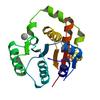
X-ray diffraction data for the Crystal Structure of Tyrosine-tRNA Synthetase from Acinetobacter baumannii with bound L-Tyrosine
First author:
D.M. Dranow
Resolution: 2.30 Å
R/Rfree: 0.18/0.23
Resolution: 2.30 Å
R/Rfree: 0.18/0.23
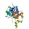
X-ray diffraction data for the Crystal Structure of Tyrosine-tRNA Synthetase from Acinetobacter baumannii
First author:
D.M. Dranow
Resolution: 2.10 Å
R/Rfree: 0.19/0.23
Resolution: 2.10 Å
R/Rfree: 0.19/0.23
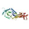
X-ray diffraction data for the Crystal Structure of Glutamate-tRNA Synthetase from Helicobacter pylori
First author:
D.M. Dranow
Resolution: 2.50 Å
R/Rfree: 0.23/0.28
Resolution: 2.50 Å
R/Rfree: 0.23/0.28
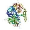
X-ray diffraction data for the Crystal structure of cytoplasmic Serine-tRNA ligase from Naegleria fowleri in complex with AMP

First author:
J. Abendroth
Resolution: 2.10 Å
R/Rfree: 0.16/0.19
Resolution: 2.10 Å
R/Rfree: 0.16/0.19
X-ray diffraction data for the Crystal structure of Tyrosine-tRNA ligase from Helicobacter pylori G27

First author:
J. Abendroth
Resolution: 2.20 Å
R/Rfree: 0.18/0.21
Resolution: 2.20 Å
R/Rfree: 0.18/0.21
X-ray diffraction data for the 2.22 Angstrom Crystal Structure of N-terminal Fragment (residues 1-419) of Elongation Factor G from Legionella pneumophila.
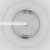
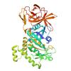
X-ray diffraction data for the 2.6 Angstrom Resolution Crystal Structure of Penicillin-Binding Protein 1A from Haemophilus influenzae
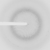
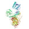
X-ray diffraction data for the Crystal structure of a GNAT Superfamily PA3944 acetyltransferase in complex with CoA (P1 space group)
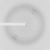
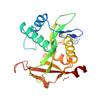
X-ray diffraction data for the A 2.05A X-Ray Structureof A Bacterial Extracellular Solute-binding Protein, family 5 for Bacillus anthracis str. Ames