120 results
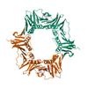
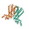
X-ray diffraction data for the CRYSTAL STRUCTURE OF A PUTATIVE DNA DAMAGE-INDUCIBLE PROTEIN (CHU_0679) FROM CYTOPHAGA HUTCHINSONII ATCC 33406 AT 1.50 A RESOLUTION
First author:
Joint Center for Structural Genomics (JCSG)
Resolution: 1.50 Å
R/Rfree: 0.16/0.19
Resolution: 1.50 Å
R/Rfree: 0.16/0.19
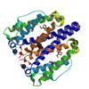
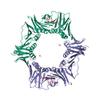
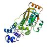
X-ray diffraction data for the Crystal structure of a Putative DNA replication regulator Hda (Sama_1916) from SHEWANELLA AMAZONENSIS SB2B at 3.00 A resolution
First author:
JOINT CENTER FOR STRUCTURAL GENOMICS (JCSG)
Resolution: 3.00 Å
R/Rfree: 0.22/0.25
Resolution: 3.00 Å
R/Rfree: 0.22/0.25
X-ray diffraction data for the Structure of DNA polymerase III subunit beta from Rickettsia conorii in complex with a natural product
First author:
Seattle Structural Genomics Center for Infectious Disease (SSGCID)
Resolution: 2.25 Å
R/Rfree: 0.18/0.23
Resolution: 2.25 Å
R/Rfree: 0.18/0.23
X-ray diffraction data for the Structure of DNA polymerase III subunit beta from Rickettsia typhi in complex with a natural product
First author:
Seattle Structural Genomics Center for Infectious Disease (SSGCID)
Resolution: 1.85 Å
R/Rfree: 0.17/0.21
Resolution: 1.85 Å
R/Rfree: 0.17/0.21
X-ray diffraction data for the Crystal structure of ferritin:DNA-binding protein DPS from Brucella Melitensis
First author:
Seattle Structural Genomics Center for Infectious Disease (SSGCID)
Resolution: 1.70 Å
R/Rfree: 0.14/0.18
Resolution: 1.70 Å
R/Rfree: 0.14/0.18
X-ray diffraction data for the The crystal structure of DNA starvation/stationary phase protection protein Dps from Yersinia pestis KIM 10
First author:
K. Tan
Gene name: dps
Resolution: 2.75 Å
R/Rfree: 0.18/0.27
Gene name: dps
Resolution: 2.75 Å
R/Rfree: 0.18/0.27
X-ray diffraction data for the 2.17 Angstrom Crystal Structure of DNA-directed RNA Polymerase Subunit Alpha from Campylobacter jejuni.
First author:
G. Minasov
Gene name: rpoA
Resolution: 2.17 Å
R/Rfree: 0.17/0.23
Gene name: rpoA
Resolution: 2.17 Å
R/Rfree: 0.17/0.23
X-ray diffraction data for the DNA-binding transcriptional repressor AcrR from Salmonella typhimurium.
First author:
J. Osipiuk
Gene name: acrR
Resolution: 1.56 Å
R/Rfree: 0.15/0.20
Gene name: acrR
Resolution: 1.56 Å
R/Rfree: 0.15/0.20
X-ray diffraction data for the Crystal structure of a PAS and DNA binding domain containing protein (Caur_2278) from CHLOROFLEXUS AURANTIACUS J-10-FL at 2.30 A resolution
First author:
Q. Xu
Resolution: 2.30 Å
R/Rfree: 0.17/0.21
Resolution: 2.30 Å
R/Rfree: 0.17/0.21
X-ray diffraction data for the Crystal structure of a predicted dna-binding transcriptional regulator (saro_1072) from novosphingobium aromaticivorans dsm at 2.10 A resolution
First author:
Joint Center for Structural Genomics (JCSG)
Resolution: 2.10 Å
R/Rfree: 0.17/0.20
Resolution: 2.10 Å
R/Rfree: 0.17/0.20