1160 results
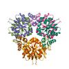
X-ray diffraction data for the Phosphopantetheine adenylyltransferase from Yersinia pestis.
First author:
J. Osipiuk
Gene name: coaD
Resolution: 2.16 Å
R/Rfree: 0.18/0.21
Gene name: coaD
Resolution: 2.16 Å
R/Rfree: 0.18/0.21
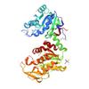
X-ray diffraction data for the Crystal Structure of Phosphoglycerate Kinase from Bacillus Anthracis
First author:
M.Chruszcz H.Zheng
Resolution: 1.68 Å
R/Rfree: 0.18/0.21
Resolution: 1.68 Å
R/Rfree: 0.18/0.21
X-ray diffraction data for the Crystal Structure of Glutaredoxin 1 from Francisella tularensis Complexed with Cacodylate

First author:
N. Maltseva
Gene name: grxA
Resolution: 2.05 Å
R/Rfree: 0.17/0.22
Gene name: grxA
Resolution: 2.05 Å
R/Rfree: 0.17/0.22
X-ray diffraction data for the Crystal Structure of Phosphoglycerate Kinase from Bacillus Anthracis
First author:
H. Zheng
Resolution: 1.68 Å
R/Rfree: 0.17/0.21
Resolution: 1.68 Å
R/Rfree: 0.17/0.21
X-ray diffraction data for the Crystal structure of bromoperoxidase from Bacillus anthracis
First author:
J. Osipiuk
Resolution: 1.74 Å
R/Rfree: 0.16/0.19
Resolution: 1.74 Å
R/Rfree: 0.16/0.19
X-ray diffraction data for the Crystal structure of 3-keto-L-gulonate-6-phosphate decarboxylase from Vibrio cholerae O1 biovar El Tor str. N16961

First author:
B. Nocek
Gene name: ulaD
Resolution: 2.10 Å
R/Rfree: 0.18/0.21
Gene name: ulaD
Resolution: 2.10 Å
R/Rfree: 0.18/0.21
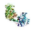
X-ray diffraction data for the Crystal Structure of the Flavohem-like-FAD/NAD Binding Domain of Nitric Oxide Dioxygenase from Vibrio cholerae O1 biovar El Tor
First author:
Y. Kim
Resolution: 2.20 Å
R/Rfree: 0.17/0.22
Resolution: 2.20 Å
R/Rfree: 0.17/0.22
X-ray diffraction data for the Crystal structure of beta-ketoacyl-acyl carrier protein reductase (FabG)(Q152A) from Vibrio cholerae

First author:
J. Hou
Gene name: fabG
Resolution: 2.55 Å
R/Rfree: 0.21/0.25
Gene name: fabG
Resolution: 2.55 Å
R/Rfree: 0.21/0.25
X-ray diffraction data for the Crystal Structure of 2-C-Methyl-D-Erythritol 2,4-Cyclodiphosphate Synthase from Francisella tularensis
First author:
Y. Kim
Gene name: ispF
Resolution: 2.65 Å
R/Rfree: 0.17/0.23
Gene name: ispF
Resolution: 2.65 Å
R/Rfree: 0.17/0.23
X-ray diffraction data for the GBAA_1210 protein, a putative adenylate cyclase, from Bacillus anthracis in complex with AMP

First author:
J. Osipiuk
Resolution: 2.10 Å
R/Rfree: 0.19/0.25
Resolution: 2.10 Å
R/Rfree: 0.19/0.25