1160 results
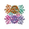
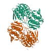
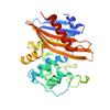
X-ray diffraction data for the Structure of putative lactate dehydrogenase from Francisella tularensis subsp. tularensis SCHU S4
First author:
T. Osinski
Gene name: mdh
Resolution: 2.20 Å
R/Rfree: 0.18/0.23
Gene name: mdh
Resolution: 2.20 Å
R/Rfree: 0.18/0.23
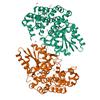
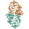
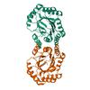
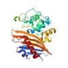
X-ray diffraction data for the Crystal structure of the beta-keto-acyl carrier protein synthase II (lmo2201) from Listeria monocytogenes
First author:
S.M. Anderson
Gene name: -
Resolution: 1.85 Å
R/Rfree: 0.15/0.18
Gene name: -
Resolution: 1.85 Å
R/Rfree: 0.15/0.18
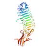
X-ray diffraction data for the 1.95 Angstrom Crystal Structure of CocE/NonD family hydrolase (SACOL2612) from Staphylococcus aureus
First author:
T. Osinski
Resolution: 1.95 Å
R/Rfree: 0.14/0.18
Resolution: 1.95 Å
R/Rfree: 0.14/0.18
X-ray diffraction data for the 2.25 Angstrom resolution crystal structure of a thymidylate kinase (tmk) from Vibrio cholerae O1 biovar eltor str. N16961 in complex with thymidine
First author:
A.S. Halavaty
Gene name: tmk
Resolution: 2.25 Å
R/Rfree: 0.21/0.26
Gene name: tmk
Resolution: 2.25 Å
R/Rfree: 0.21/0.26
X-ray diffraction data for the 1.8 Angstrom resolution crystal structure of orotidine 5'-phosphate decarboxylase (pyrF) from Campylobacter jejuni subsp. jejuni NCTC 11168

First author:
A.S. Halavaty
Gene name: pyrF
Resolution: 1.80 Å
R/Rfree: 0.17/0.20
Gene name: pyrF
Resolution: 1.80 Å
R/Rfree: 0.17/0.20
X-ray diffraction data for the Crystal structure of anabolic ornithine carbamoyltransferase from Bacillus anthracis in complex with carbamoyl phosphate and L-norvaline
First author:
I.G. Shabalin
Gene name: argF
Resolution: 1.74 Å
R/Rfree: 0.15/0.17
Gene name: argF
Resolution: 1.74 Å
R/Rfree: 0.15/0.17
X-ray diffraction data for the 1.1 Angstrom Crystal Structure of Putative Modulator of Drug Activity (MdaB) from Yersinia pestis CO92.
First author:
G. Minasov
Gene name: mdaB
Resolution: 1.10 Å
R/Rfree: 0.10/0.12
Gene name: mdaB
Resolution: 1.10 Å
R/Rfree: 0.10/0.12
X-ray diffraction data for the Crystal structure of the double mutant (S112A, H303A) of B.anthracis mycrocine immunity protein (MccF)

First author:
B. Nocek
Resolution: 1.40 Å
R/Rfree: 0.11/0.15
Resolution: 1.40 Å
R/Rfree: 0.11/0.15
X-ray diffraction data for the The structure of a sensor domain of a histidine kinase from Vibrio cholerae O1 biovar eltor str. N16961
First author:
K. Tan
Resolution: 1.98 Å
R/Rfree: 0.18/0.20
Resolution: 1.98 Å
R/Rfree: 0.18/0.20
X-ray diffraction data for the The crystal structure of phosphoribosylglycinamide formyltransferase from Streptococcus pneumoniae TIGR4
First author:
K. Tan
Resolution: 2.70 Å
R/Rfree: 0.20/0.28
Resolution: 2.70 Å
R/Rfree: 0.20/0.28
X-ray diffraction data for the Crystal structure of the 3-dehydroquinate synthase (DHQS) domain of Aro1 from Candida albicans SC5314 in complex with NADH
X-ray diffraction data for the 1.75 Angstrom Resolution Crystal Structure of D-alanyl-D-alanine Endopeptidase from Enterobacter cloacae in Complex with Covalently Bound Boronic Acid
X-ray diffraction data for the 1.05 Angstrom Resolution Crystal Structure of UDP-N-acetylglucosamine 1-carboxyvinyltransferase from Acinetobacter baumannii in Covalently Bound Complex with (2R)-2-(phosphonooxy)propanoic Acid.
X-ray diffraction data for the Crystal structure of a GNAT Superfamily PA3944 acetyltransferase in complex with CoA (P1 space group)
X-ray diffraction data for the A 2.05A X-Ray Structureof A Bacterial Extracellular Solute-binding Protein, family 5 for Bacillus anthracis str. Ames
X-ray diffraction data for the Listeria monocytogenes internalin-like protein lmo2027
X-ray diffraction data for the 1.25 Angstrom Crystal Structure of Chitinase from Bacillus anthracis.
X-ray diffraction data for the 1.0 Angstrom Crystal Structure of pre-Peptidase C-terminal Domain of Collagenase from Bacillus anthracis.
X-ray diffraction data for the 2.2 Angstrom Crystal Structure of ABC Transporter Substrate Binding Protein CtaP (Lmo0135) from Listeria monocytogenes.

X-ray diffraction data for the 1.35 Angstrom Crystal Structure of C-terminal Domain of Glycosyl Transferase Group 1 Family Protein (LpcC) from Francisella tularensis.
X-ray diffraction data for the 2.11 Angstrom resolution crystal structure of betaine aldehyde dehydrogenase (betB) P449M/Y450L double mutant from Staphylococcus aureus in complex with NAD+ and BME-modified Cys289
X-ray diffraction data for the 1.8 Angstrom Crystal Structure of the 3-Dehydroquinate Dehydratase (aroD) from Salmonella typhimurium LT2 with Nickel Bound at Active Site
X-ray diffraction data for the 1.3 Angstrom Resolution Crystal Structure of UDP-N-acetylglucosamine 1-carboxyvinyltransferase from Streptococcus pneumoniae in Complex with (2R)-2-(phosphonooxy)propanoic acid.
X-ray diffraction data for the Crystal Structure of K83A Mutant of Class D beta-lactamase from Clostridium difficile 630
First author:
G. Minasov
Gene name:
Resolution: 1.88 Å
R/Rfree: 0.17/0.20
Gene name:
Resolution: 1.88 Å
R/Rfree: 0.17/0.20
X-ray diffraction data for the Crystal Structure of C79A Mutant of Class D beta-lactamase from Clostridium difficile 630
First author:
G. Minasov
Gene name:
Resolution: 1.95 Å
R/Rfree: 0.18/0.21
Gene name:
Resolution: 1.95 Å
R/Rfree: 0.18/0.21